The Floor Of The Cranium Is Primarily Formed By The

Crus Cerebri Google Search Brain Anatomy And Function Brain Stem Brain Anatomy

Surface Markings Of The Abdomen Human Anatomy Abdomen Anatomy Abdominal

11 6 Axial Muscles Are Muscles Of The Head And Neck Vertebral Column Trunk And Pelvic Floor Head Muscles Temporomandibular Joint Muscle

Anatomy Of The Pudendal Nerve Health Organization For Pudendal Education Pelvis Anatomy Pelvic Bone Anatomy Bones

Anatomy Of The Pudendal Nerve Health Organization For Pudendal Education Vulvodynia Neuralgia Nerve Health

Anterior Cranial Fossa Nasal Cavity And Paranasal Sinuses Radiology Key
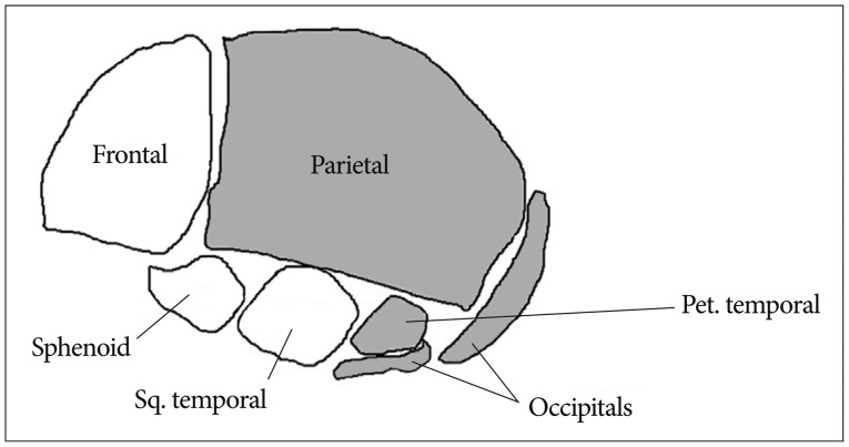
The two bones which primarily make up the walls of the calvarium are the.
The floor of the cranium is primarily formed by the.

The Renal Corpuscle Anatomy And Physiology Physiology Renal

7 5 Embryonic Development Of The Axial Skeleton Anatomy And Physiology
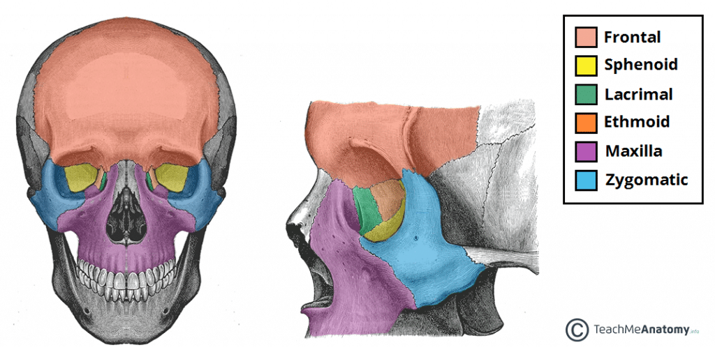
The Bony Orbit Borders Contents Fractures Teachmeanatomy

The Sensitivity Of This Stretch Receptor Is Adjusted By Gamma Motor Neurons Which Innervate These Contractile Motor Neuron Massage Human Anatomy And Physiology
Source : pinterest.com
